Aortic Arch
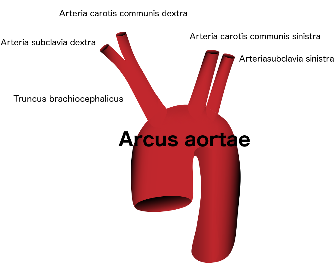
An arched highest part of aorta situated between the ascending and descending parts of the aorta. From the aortic arch spring the large arterys for the head and the arms.
The usual aortic arch shows only the
Exit of Truncus brachiocephalicus (supplies the carotis communis dextra and subclavia dextra arteries),
then follow
the "Arteria carotis communis sinistra" and
the "Arteria subclavia sinistra".
Normal aortic arc
Anomalies of aortic arch
Approximately 26% of the patients have anomalies of the aortic arch that are not symptomatic and are only diagnosed during imaging.
However, knowledge of this can be important in the context of surgical and endovascular therapy. About 60 % of the cases of aortic dissection are located in the aortic arch.
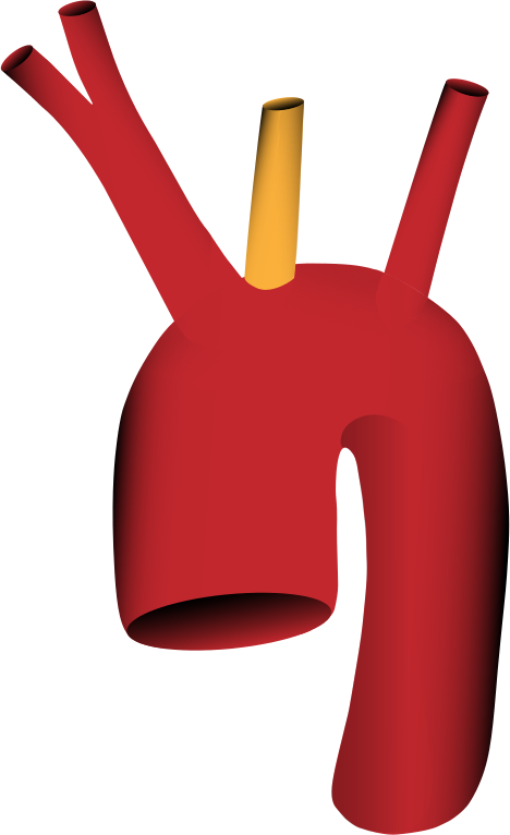
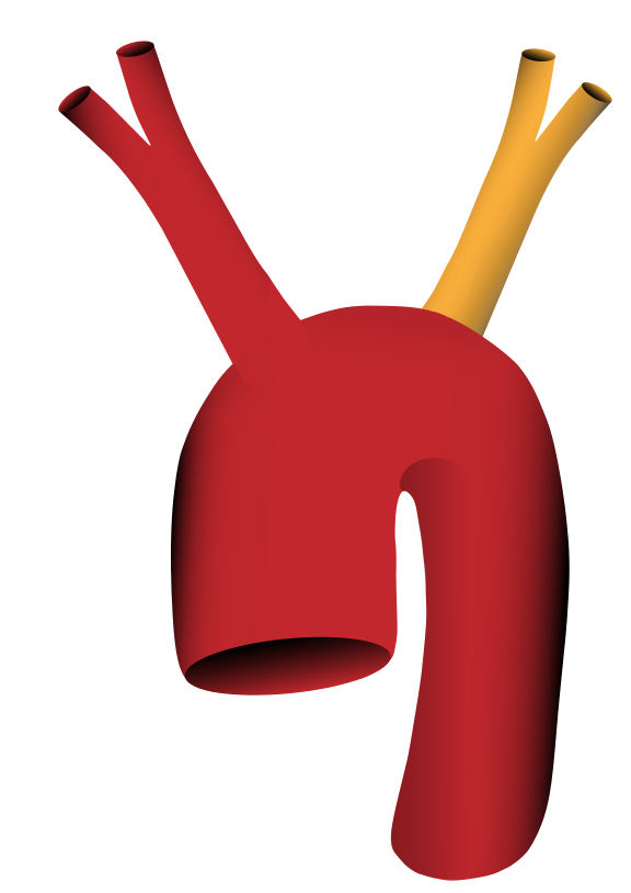
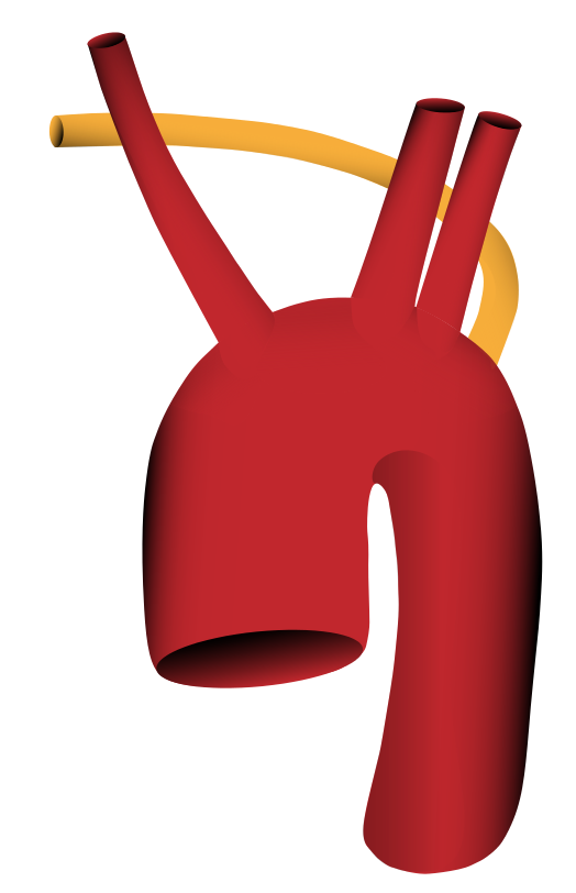
Here you can see examples of anomalies of the aortic arch (according to Lippert and Pabst).
Truncus brachiocephalicus (with Arteria subclavia dextra and Arteria carotis communis dextra) AND the Arteria carotis communis sinistra arise together from the aortic arch (in 13% of the cases)
In addition to the subclavian dextra artery and the carotis communis dextra artery, the carotis communis sinistra artery is also derived from the truncus brachiocephalicus (in 9% of cases).
There are two Trunci brachiocephalici, each with the Arteria subclavia dextra and Arteria carotis communis dextra and with the Arteria subclavia sinistra and Arteria carotis communis sinistra (in 1% of cases)
The arteria subclavia dextra originates only after the arteria subclavia sinistra from the aortic arch
Aortic arch with separate outlet of left arteria vertebralis


